MYRIAN XP-ONCOLOGY
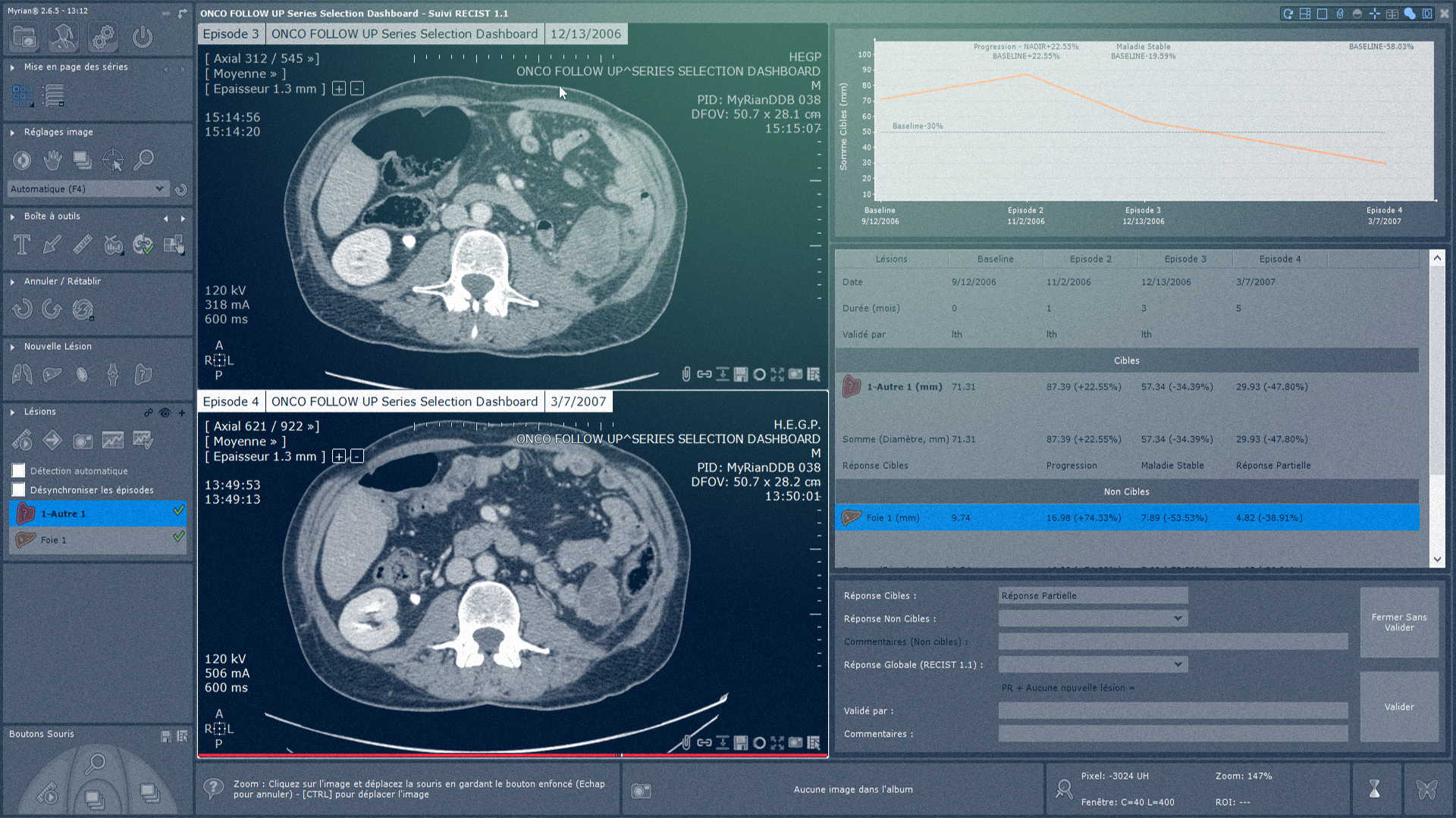
Optimize multi-modality oncology follow-up with innovative analysis, interpretation and reporting tools. Easily and intuitively assess therapeutic response at different measurement points. Innovative tools and a structured workflow dedicated to oncology follow-up.
Compatible with CT, MRI and PET-CT
Automatic segmentation and measurement of lesions in a single click
Automatic selection of patient's comparative examinations
Easy lesion tracking
Automatic synchronization of images from different examinations
Support for standard evaluation criteria (RECIST 1.1) and customized evaluation criteria
Summary dashboard for oncology follow-up
Management of different measurement points (Baseline, NADIR)
MYRIAN XP-LIVER
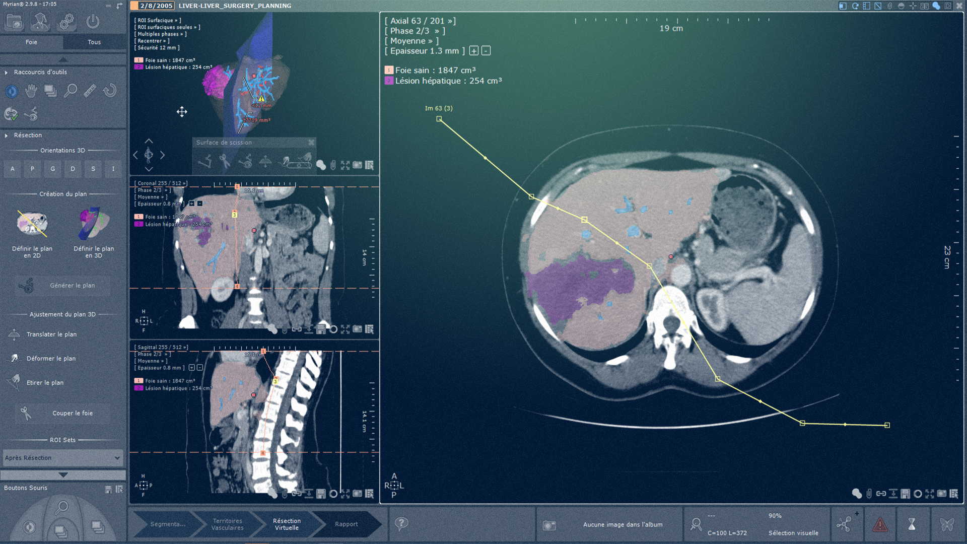
A complete solution dedicated to liver lesion detection and virtual hepatectomy. Benefit from semi-automatic segmentation of the liver, lesions and vascular territory.
Semi-automatic segmentation of liver and anatomical structures (parenchyma, lesions, vascular trees)
Visualization of segmented anatomical structures
One-click calculation of segment volume
Creation and optimization of the resection surface in 2D MPR and 3D representation of the liver model
Surgical planning of hepatectomy: 3D simulation of the cutting plane, automatic calculation of healthy and resected liver volumes
Creation of 3D PDF reports
MYRIAN XP-COLON
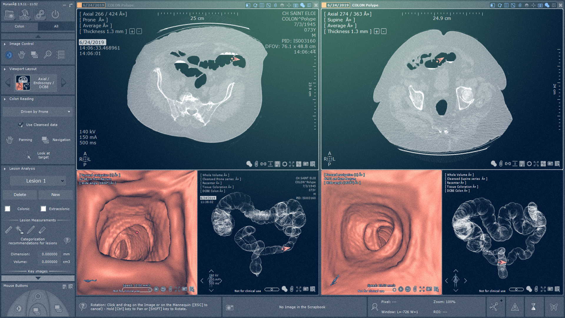
Visualize your colon examinations in 3D thanks to automatic segmentation, and navigate in fully synchronized mode. Use multiple automatic tools to analyze your exam in just a few clicks and focus on your diagnosis.
Dedicated visualization screens: endoscopy, colon DCBE, Fullsight, 3D polyp zoom
Automatic colon segmentation
Measurement and localization of polyps
Automatic calculation of distance from lesion to rectum
Virtual endoscope navigation and 3D ribbon view
Generation of a complete fat assessment report
Polyp evaluation and reporting according to C-RADS guidelines
Structured workflow dedicated to your examination

MYRIAN XP-ABDOFAT
Benefit from a dedicated protocol for automatic quantification and segmentation of abdominal adipose tissue.
One-click segmentation of peripheral and visceral adipose regions
Automatic calculation of adipose ratio in peripheral and visceral areas
Automatic calculation of waist circumference
Setting of fat intensity threshold
Manual tools to modify segmentation
Generation of a complete fat assessment report