MYRIAN XP-LUNG NODULE
Use a set of advanced tools to segment, quantify and follow-up lung nodules intuitively. Simplify screening and therapeutic follow-up.
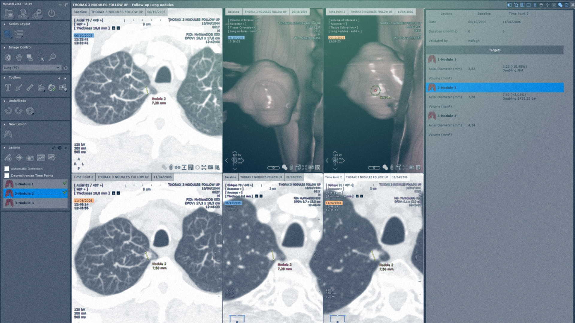
Semi-automatic lung nodule segmentation
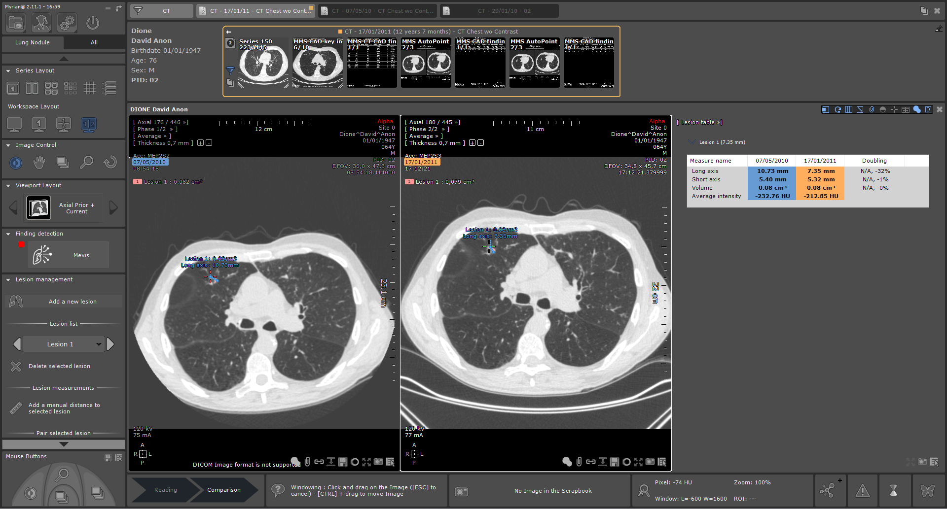
Simultaneous review of history
Automatic synchronization of examinations
Longitudinal tracking of nodules
Automatic calculation of doubling time and growth percentage
3D visualization of lesions
MYRIAN XP-LUNG NODULE OPTION IA
Choose an AI algorithm perfectly integrated into your workflow to automatically detect nodules and track their evolution.
Automatic detection of solid and sub-solid lung nodules thanks to artificial intelligence
Synchronized comparison of anomalies detected on two examinations
One of the highest-performing solutions on the market (92.4% sensitivity, including on ground-glass nodules)
MYRIAN XP-LUNG
A complete solution for your lung examinations: lung tissue segmentation, 3D image visualization, parenchyma and airway analysis. Facilitate and secure your imaging analyses with high-performance functionalities.
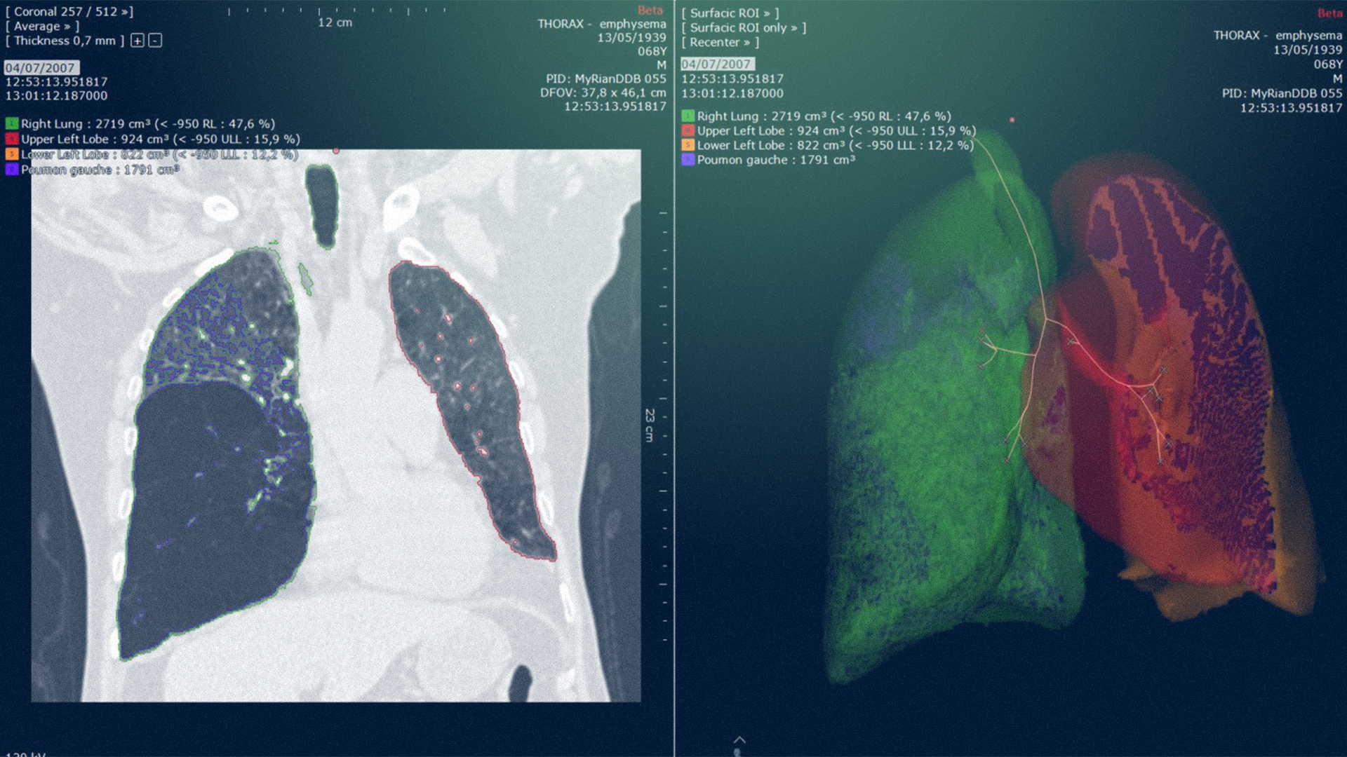
Segmentation and quantification of low-attenuation tissues
Segmentation and quantification of tissues affected by Covid-19
Automatic segmentation of lung volumes and airways
Airway visualization for CPR and stenting as part of surgical planning
Segmentation des lésions
3D visualization of interactions between vasculature, airways and lesions
Lobectomy simulation with automatic calculation of post-procedure lung volumes