MYRIAN XM-MG
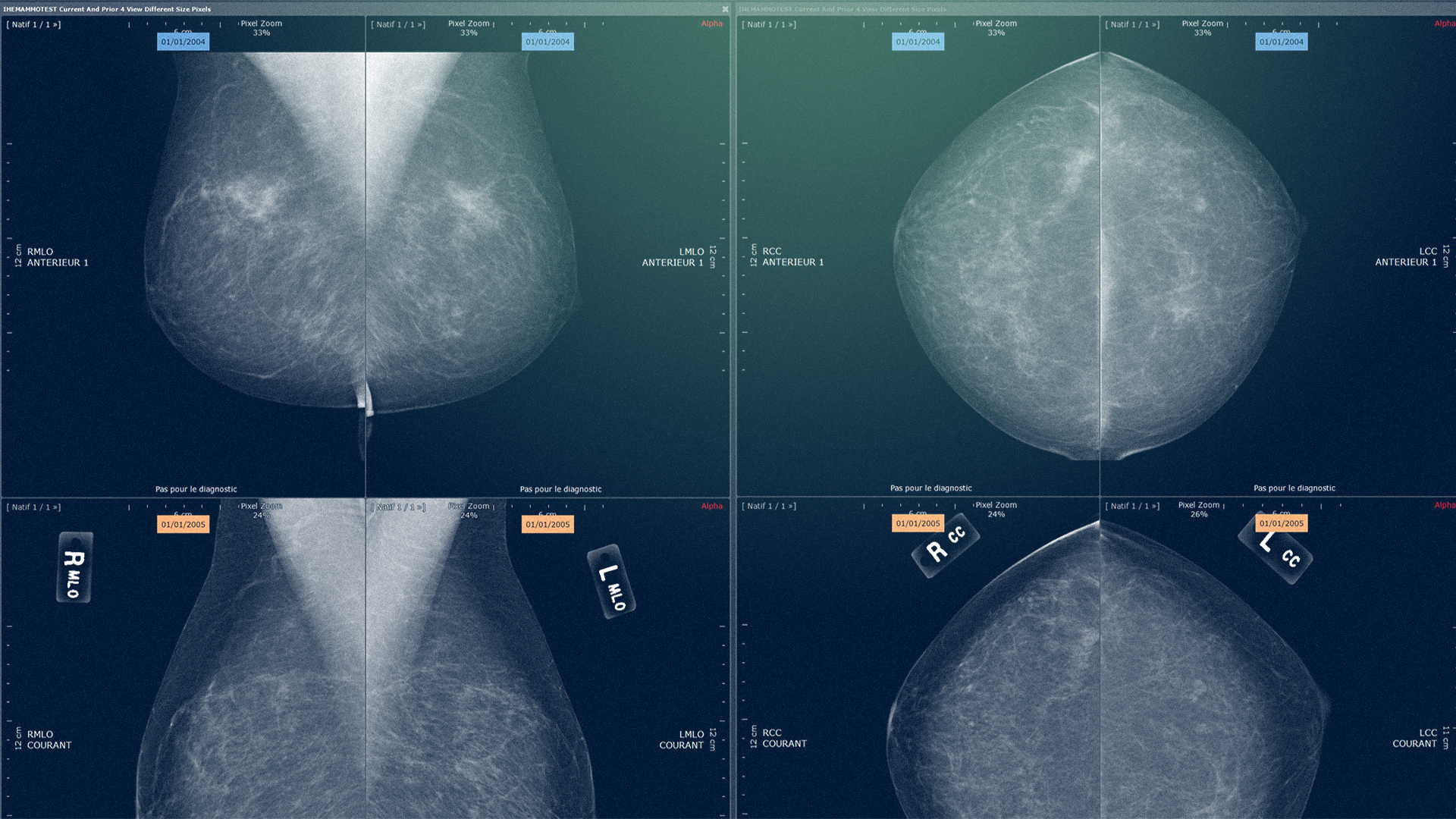
Benefit from a dedicated workflow and all the tools you need to analyze your mammographic examinations, whatever your clinical habits (standard or mirrored visualization, 2D imaging with or without tomosynthesis).
Multimodality MRI and ultrasound visualization
Optimized display for mammography and tomosynthesis examinations
Display adapted for comparative examinations
1:1 viewing mode with intuitive navigation
Planning tool with localizer
Tomosynthesis display with synthetic image
Adjustable, synchronized tomosynthesis magnifier with volume navigation
One-click display switching between synthetic and mammographic images

MYRIAN XP-BREAST
Discover a working environment entirely dedicated to breast MRI. Optimize your workflow and boost your productivity: customizable display protocols and cutting-edge diagnostic tools to enhance the quality of your patients' care.
Comprehensive range of multi-phase and multi-volume tools
Simplified navigation through the different acquisition phases
Comprehensive parametric maps to help detect lesions
Calculation of subcontracted series with or without motion correction
Lesion measurements
Plotting of breast lesions on a sector map
Automatic transcription of lesion measurements (size, volume, ADC) in the report
Structured report including lesion scoring according to ACR score
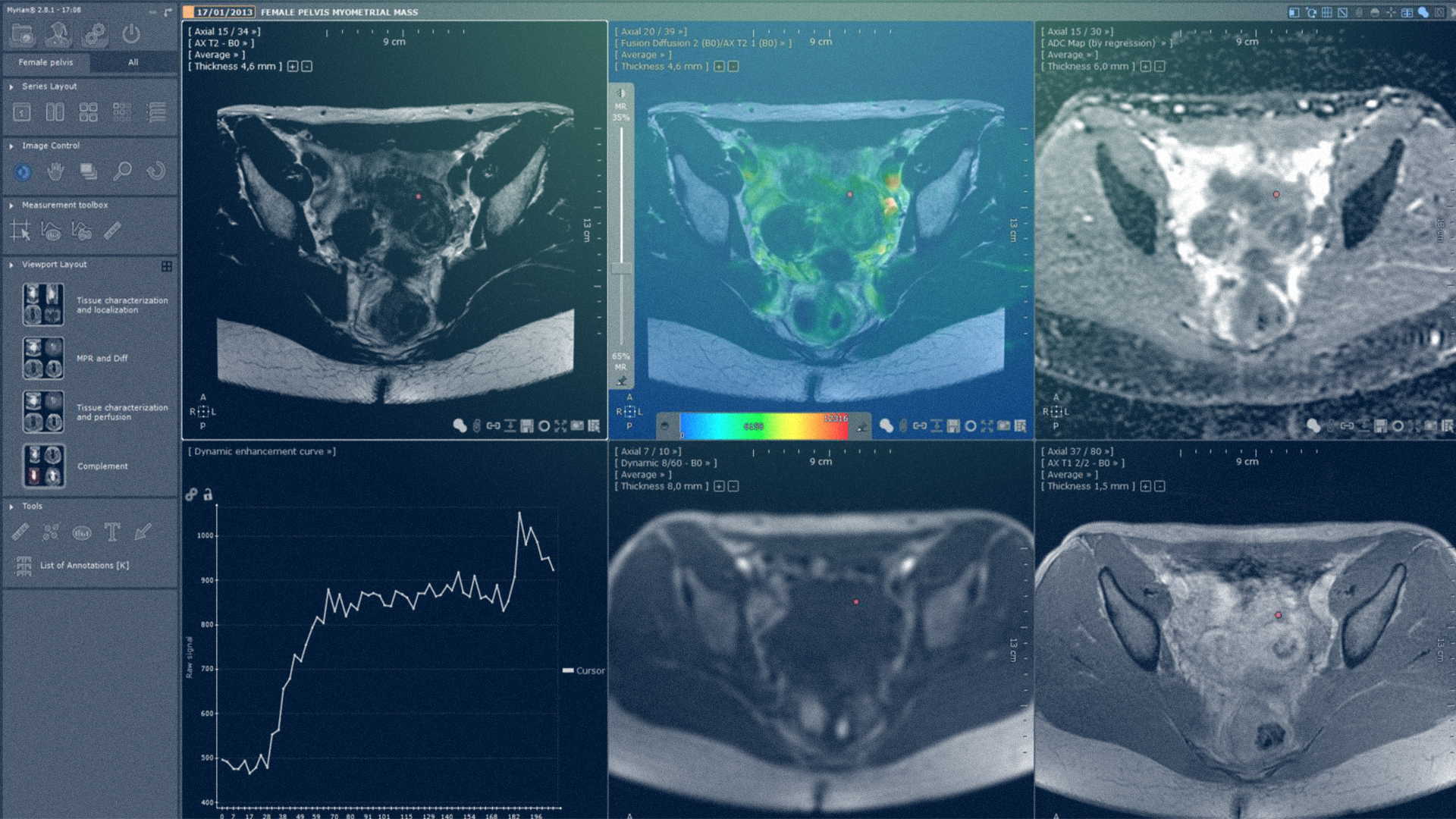
MYRIAN XP-FEMALE PELVIS
Full clinical functionality for optimized visualization and analysis of MRI scans of the female pelvis. A set of applications and tools dedicated to the grading of :
Ovarian masses
Cervical lesions
Myometrial masses
Endometrial lesions
Ectopic endometrial tissue lesions
Simplified navigation through the different acquisition phases
Customizable display of dynamic series, ADC, diffusion, subtractions
Structured report including automatic calculation of O-RADS score