A 2-fold increase
in CT examination volumes in 15 years 1
Limited interpretation time
Risk of error estimated at 10%2
The first automated solution for CT-TAP interpretation, Liflow enables faster, more complete analysis of oncology imaging examinations.
Added values Liflow
Facilitates comparative reading
Simplify the analysis of your oncology follow-up examinations with Liflow: save time and automate reading by anatomical region.
Optimize lesion detection
With Liflow, a multi-organ AI-enriched solution, obtain detection results on a single image as soon as the examination is opened, benefit from automatic measurements and interact directly with the results, in a single interface.
Automate oncology follow-up
Liflow makes it quick and easy to follow up your patient: compare lesions, find antecedents, automatically calculate tumor evolution and RECIST 1.1 score.
Integrated into your radiology workflow
With Liflow, open your images from PACS and retrieve relevant patient history. View multi-organ detection results as soon as you open the exam, directly in our viewer.
Multi-organ AI to optimize lesion detection and follow-up
AI for lung lesions
Developed by MeVis Medical Solutions AG
> Automatic detection of lung lesions
> Results available as soon as the examination is opened
> Interaction with result possible
> 92.4% sensitivity for solid, subsolid and calcified nodules3
IA DEDICATED TO LIVER
Developed with Guerbet
> Automatically detects and segments lesions
> Automatic measurements: long axis, short axis, volume, density
> Results available as soon as the examination is opened
> Interaction with results possible
> Sensitivity of 75% (average radiologist sensitivity: 61.7%) 4
Liflow Innovation in oncology imaging
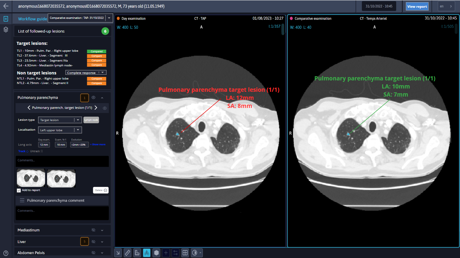
Better interpret your oncology imaging examinations
Liflow automates lesion identification and measurement, providing you with all the tools you need for each anatomical zone.
The results of multi-organ artificial intelligence are displayed on the same image, making it easier for you to take measurements and help you with your diagnosis.
Facilitate your oncology follow-up with automatic calculation of tumor evolution and RECIST 1.1 score, directly integrated into a structured report.
Integrated into the radiology workflow, Liflow provides access to anteriorities and the display of different time points, for a 360° view of the patient.
A cloud-based solution, Liflow encourages optimized collaboration between clinicians in the multidisciplinary management of cancer patients.
To help you analyze your exams :
> Interpretation guide to facilitate your analysis
> Native AI integration to detect suspicious lesions
> Automatic RECIST 1.1 follow-up for more accurate oncology assessment
Ready to experience Liflow?
TEST THE SOLUTION
A solution backed by recognized experts in the fight against cancer
Liflow has been developed in collaboration with radiologists from Cancer Research Centers and University Hospitals.
Want to know everything there is to know about using the Liflow solution and its AIs? Our user documentation will answer all your questions!
Please fill in this form to receive the link to download the resource:
Your questions answered
How does Liflow simplify my oncology diagnosis and follow-up?
Liflow automates lesion detection and measurement. Its workflow, specially designed for oncology follow-up, saves the radiologist clicks, allowing him or her to benefit from windowing and tools adapted to each anatomical zone, and to concentrate on clinical interpretation. Automatic integration of tumor progression and RECIST 1.1 indicator facilitate examination analysis, enabling more accurate assessment of patient data. In addition, Liflow facilitates reporting and ensures longitudinal structuring of follow-up data, simplifying the monitoring and evolution of oncological pathologies from one examination to the next.
How is AI integrated into the Liflow workflow?
In Liflow, multi-organ AI performs image pre-processing, enabling the automatic identification, retrieval and analysis of examinations relevant to oncology follow-up. The detection results generated by the AI are directly displayed as soon as the examination is opened, facilitating the interpretation and measurement process. Liflow offers interaction with the results, enabling professionals to use them directly in their oncology follow-up practice.
Want to learn more ?
Contact our team
1 Between 2009 and2020, Bruls and al. "Workload for radiologists during on-call hours : dramatic increase in the past 15 years". Insights Imaging 11, 121 (2020)
2 Kasalak, Ömer and al. "Work overload and diagnostic errors in radiology". European journal of radiology vol. 167 (2023)
3 In-house study MeVis Medical Solutions AG Veolity
4 Phase II study by Kyoto University. Publication Eur. Jour. Rad. June 2023
CONTACT DETAILS
1231 Avenue du Mondial 98, 34000 Montpellier, France