Elevate your ability to detect suspicious lesions with DUOnco™, a comprehensive suite of standalone AI solutions co-developed with Guerbet.
Our advanced AI technology automates lesion detection, delivering critical insights to enable faster and more accurate clinical decision-making.
Automatic detection of suspicious lesions
Streamline your analysis with advanced AI-powered lesion detection. Automate complex measurements and leverage fast, precise CT image analysis to enhance your diagnostic accuracy.
Seamless data integration
Access detected lesions directly within your PACS or DICOM viewer via DICOM SC, DICOM GSPS, DICOM SR and DICOM SEG, ensuring critical data is at your fingertips.
Cost efficiency for healthcare providers
Minimize costly late-stage treatments through early, accurate lesion detection and faster interpretation times.
Our AI solutions dedicated to oncology
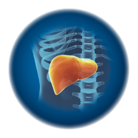
DUOnco™ Liver
AI that detects even the most subtle liver damage, long before it becomes obvious.
The liver, the primary site of metastasis for many cancers, remains one of the most complex organs to analyze. Between steatosis, cirrhosis, and tumor heterogeneity, image interpretation varies greatly between readers, making early diagnosis particularly difficult.¹
> Automatic lesion detection and segmentation
> Automatic lesion measurements and quantification (diameters, volume, average Hounsfield units)
> Reduced inter-operator variability
DUOnco™ Liver improves efficiency and reduces the risk of error by delivering enhanced exactness in liver exam analysis: 94% sensitivity (+52% compared to average radiologist sensitivity²)
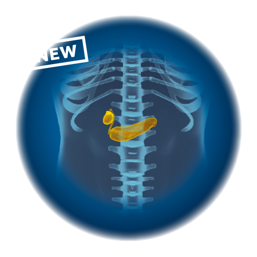
DUOnco™ Pancreas
Your AI-powered assistant that detects small lesions at an early stage
Pancreatic cancer is among the most difficult cancers to detect early, with no reliable early-stage screening methods. When identified at <2 cm, the five-year survival rate can rise from 9% to 30%.³
> Automatic lesion detection and segmentation of all lesion types and sizes
> Automatic main pancreatic duct detection and segmentation
> Three-level priority grouping for findings
> AI processing status on your worklist
DUOnco™ Pancreas is designed to help radiologists identify subtle pancreatic lesions earlier, improving the chances of better patient outcomes: 93,6% sensitivity
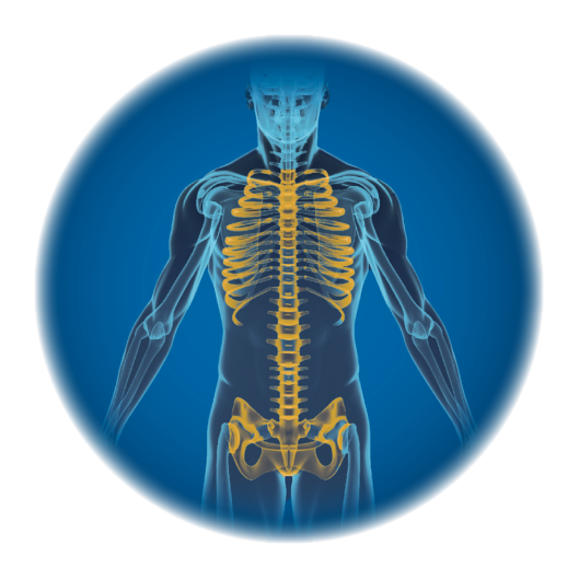
DUOnco™ Bone
The World’s first CE-marked AI algorithm for the detection of bone lesions in CT-TAP!
Nearly 30% of bone lesions go undetected in CT scans⁴. Due to the heterogeneity of bone tissue and lesions, and its extensive distribution throughout the body, bone has the highest diagnostic error rate of any organ on CT⁵.
> Automatic detection of bone lesions (including primary lesions and metastases)
> Automatic lesions localization through 3D bounding boxes
> Automatic lesion detection and segmentation of all lesion types and sizes
DUOnco™ Bone enhances accuracy to support clinical assessment, streamlining your interpretation of oncologic imaging: 73% sensitivity for lesion > 1cm (+22.4% compared to average radiologist sensitivity⁶)
Want to know everything about using our DUOnco™ AI solutions? The user documentation will answer your questions!
I FILL OUT THE FORM
Questions you may have
How does DUOnco™ simplify my diagnostic work?
DUOnco™ helps you keep an eye on oncology findings, without adding time to your workflow. It automatically detects suspicious lesions, even small ones that may be overlooked, and highlights them directly in your PACS or DICOM viewer for fast, targeted review. By automating calculations and converting visual data into standardized measurements, DUOnco™ saves time, reduces manual workload, and supports more confident clinical decisions. Our AI solutions are designed for real-world oncology practice to enhance patient care.
What is the added value of DUOnco™ compared to other AI solutions?
DUOnco™ combines clinical performance, seamless integration, and flexibility. Co-developed with oncology experts, it offers accuracy and reliability you can trust. Unlike many AI tools that require workflow changes, DUOnco™ integrates effortlessly into your existing systems. With rapid, standardized results, it helps improve diagnostic quality and reduce unnecessary procedures.
What support will I receive after installation?
Our sales team will provide personal support and answer questions you may have over time. You’ll get access to training and an on-site or remote follow-up service. Our Support engineers, based in Montpellier, are available on the phone or by email from Monday to Friday for day-to-day remote support.*
* Support available via warranty year or service contract subscription
Want to learn more?
Contact our team
1. Artificial intelligence-powered software detected more than half of the liver metastases overlooked by radiologists on contrast-enhanced CT. Nakai, Hirotsugu et al. European Journal of Radiology, Volume 163.
2. Phase II study by Kyoto University (published in the European Journal of Radiology, June 2023)
3. Pongprasobchai S, Pannala R, Smyrk TC, Bamlet W, Pitchumoni S, Ougolkov A, de Andrade M, Petersen GM, Chari ST. Long-term survival and prognostic indicators in small (< or = 2 cm) pancreatic cancer. Pancreatology. 2008
4. Ha JY, Jeon KN, Bae K, Choi BH. Effect of Bone Reading CT software on radiologist performance in detecting bone metastases from breast cancer. Br J Radiol 2017; 90: 20160809
5. Kasalak Ö et al. Work overload and diagnostic errors in radiology. European Journal of Radiology 2023, Volume 167, 111032
6. For lesions ≥10 mm and <30 mm. Noguchi, S., Nishio, M., Sakamoto, R. et al. Deep learning–based algorithm improved radiologists’ performance in bone metastases detection on CT. Eur Radiol. 2022
CONTACT DETAILS
1231 Avenue du Mondial 98, 34000 Montpellier, France